Why Your MRI Looks “Normal” but You Still Have a CSF Leak with Dr. Andrew Callen
Description
Spinal CSF leaks are more common in connective tissue disorders than many realize. Dr. Andrew Callen on why scans read as normal — and what finds them.
Watch
Guests
Transcript
[00:43] Dr. Linda Bluestein: Welcome back, every bendy body, to the Bendy Bodies Podcast. I'm your host, Dr. Linda Bluestein, the Hypermobility MD, a Mayo Clinic-trained physician dedicated to helping you navigate EDS and complex chronic illness. Today I'm joined by Dr. Dacre Knight, who is not only an expert in EDS, HSD, POTS, and mast cell disorders, but is also joining me as a recurring co-host. Dr. Knight recently became the medical director of the UVA Health EDS and Hypermobility Disorders Center, which is officially partnering with Bendy Bodies. I am so excited to have this conversation today with neuroradiologist Dr. Andrew Callen.
In people with connective tissue disorders, CSF leaks, or cerebrospinal fluid leaks, are so much more common than most people realize. People can suffer for years, if not decades, before getting appropriately treated. So this is a really important conversation today.
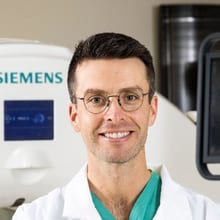
Dr. Callen is an associate professor of radiology and neurology at the University of Colorado School of Medicine and the founder and director of the CU CSF Leak Program, Colorado's first multidisciplinary center dedicated to diagnosing and treating spinal CSF leaks. A national and international leader in this field, he has authored over 60 manuscripts, including more than 30 peer-reviewed publications and 2 textbook chapters on CSF dynamics and spinal CSF leaks. He is also the inventor of a novel patient positioning device for dynamic CT myelography that improves diagnostic accuracy. Dr. Callen is a founding member of the International Spinal CSF Leak Society and serves on medical advisory boards for US and Canadian CSF leak foundations.
I'm so excited about this conversation and can't wait for you to learn from Dr. Callen. As always, this information is for educational purposes only, and it's not a substitute for personalized medical advice. Stick around until the very end so you don't miss any of our special hypermobility hacks. Here we go.
Well, I am so excited to be here with Dr. Andrew Callen. I've been wanting to talk to you for such a long time, so thank you so much for coming on Bendy Bodies.
[03:03] Dr. Andrew Callen: Thank you for having me. It's my pleasure.
[03:03] Dr. Linda Bluestein: Fantastic. So we know that CSF leaks are something that are really more common in people with connective tissue disorders. Let's just start out — if you could explain what CSF leaks are and why people can get things like spontaneous intracranial hypotension.
[03:16] Dr. Andrew Callen: Sure. Starting from basics, just a definition of terms: CSF stands for cerebrospinal fluid. It is a clear fluid — looks like water — surrounding the brain and spinal cord. It's contained by the dura, which is the sort of sacral lining that holds that fluid in. It participates not only in cushioning or a mechanical presence, but also it's very important in the metabolic function of the brain and many other processes that are being actively investigated currently.
Doctors have known for a long time that if we put a needle in someone's back, for example, to do a spinal tap, a lumbar puncture, and take fluid out, that people may get a headache and associated other symptoms. But really, a more recent understanding has emerged that these leaks can occur spontaneously. When we say spontaneously, we mean that there wasn't an obvious precipitating cause. Some people — a little less than half, around 40% of patients — will experience some sort of very minor trauma. For example, they fell down or they bumped their head. But nonetheless, we call these spontaneous rather than traumatic, which can have its own distinct appearance.
When they occur, they occur in 3 major forms. The first form is when there's a little bone spur in the spine from wear-and-tear arthritis that pokes a hole in the dura, usually in the front of the dura, and fluid comes out and we see a big fluid collection in the spine. The second is on the side of the spine. The spinal cord gives off nerve roots. Those nerve roots are branch points from the dura, and they can tear. It's these types in particular that seem to be most associated with connective tissue disease. They tend to occur in the lower thoracic spine mostly. And these, while it seems like — well, it's a hole and fluid comes out, that should be straightforward — have also undergone a renaissance in understanding, which we can get into in more detail later.
And then in 2013–2014, a third type of CSF leak was discovered: the CSF venous fistula. This is not a hole in the dura with fluid pouring out, but rather a vein — a normal vein that is participating in normal physiologic CSF resorption every day — that loses that regulatory capacity, and the fluid just gets drained through this vein unchecked. So the CSF is being lost, but into the bloodstream. There's no fluid collection. A regular myelogram where we inject X-ray dye into the spine — the test that's done in almost every hospital in the country — will not find it. You have to do a very special type of myelogram. That was part of the reason we couldn't find them.
And then, basically, this will result in the syndrome where, classically, we have what's characterized as an orthostatic headache — a headache that is worse when we are upright and better when we are flat. But that too has undergone a really big revolution in terms of understanding the symptom complex of this disease. It's really much more complicated than that. I can't tell you how many of my patients say, "Dr. Callen, it's not a headache. It's a different feeling. It's a sensation. There are other things going on with it." Some people have no headache at all. And particularly with the CSF venous fistula subtype, the presentation can be very atypical — though to me, what's atypical is really just that one basic classic story, and we're learning more and more about the different ways these patients can present. It makes it really challenging because this is not the paradigm we as doctors learn about in med school. We just say, "Oh, it's a headache," and that's it.
[07:09] Dr. Linda Bluestein: For sure. I'm an anesthesiologist, so I learned about, you know, you do a wet tap or you do a spinal for doing a subarachnoid block, and yes, you may get a CSF leak headache and then you might do a blood patch — it was a very straightforward thing. Spontaneous CSF leaks are definitely something I had not been aware of at all before. They're so often overlooked and misdiagnosed. I know that normal imaging — quote, "normal imaging" — is also a huge part of the problem. Can you explain more about why these are so often overlooked and misdiagnosed? We'll get more into what people should do if they strongly suspect they have a leak and they have normal imaging. But what kinds of symptoms should we be looking for that don't fit the classic patterns?
[07:56] Dr. Andrew Callen: There's a lot of great stuff to talk about in that question. I just want to briefly touch on the fact that we're talking about spontaneous CSF leaks today, but half my patients are people suffering from chronic doctor-caused leaks. This is another thing we've come to understand — certainly as an anesthesiologist, as a proceduralist, in neuroradiology, we were taught this is a benign self-limiting thing. Post-dural puncture headache: have them lay down, drink some caffeine, they'll be fine. We now know, by a relatively robust body of evidence, that this can become a chronic debilitating disease as well. So I feel very strongly that it requires more recognition.
But in terms of why this is overlooked — there are so many ways to look at this. The first is probably clinical recognition. If somebody does not come in and say, "I have a headache that is horrible when I stand up, it's in the back of my head, it's like a pressure sensation, and when I lay down it goes away" — if they don't say that, then their doctor probably won't be cued to think, "Could this be a CSF leak?" And if they say, "Well, when I first started I noticed that, but as it went on, when I laid down I didn't get as much relief," or they're describing vestibulocochlear symptoms — that's a very common set of symptoms that go along with a spinal CSF leak.
What I mean by that is ringing in the ears, a sense of imbalance. Patients very frequently say it's not dizziness, it's not like the room is spinning — it's almost like they feel intoxicated or like they're on a boat. People frequently describe aural fullness, meaning it feels like they need to pop their ears or they're underwater. They often have hearing issues — sensorineural hearing loss, feeling like they can't understand what people are saying to them. And along with that, cognitive symptoms — people commonly refer to it as brain fog: cognitive slowing, just feeling like they're not themselves. They can't do normal activities of daily living, balance a checkbook, talk to people without struggling to find the correct words.
These symptoms go on and on, and the problem is that they're not specific for this disease. I tell my patients: if you'd talked to me 5 years ago, I would've been much more certain about things. They would say, "I have this symptom — could that be my leak?" And I would say, "No, no, that's not a leak." And then we would treat their CSF leak and it would go away. So what do I know? There's not a specific clinical question I can ask a patient and say, "Aha, therefore it is or is not a leak." And that's why we rely so much on the imaging.
[10:59] Dr. Linda Bluestein: Yeah, that's so tricky because we know that this same population of people is at risk for Chiari malformation, dysautonomia, cranial cervical instability, so many things — elevated intracranial pressure, which is obviously the opposite. It seems like they probably get mislabeled super commonly. It would be nice if we had really highly specific things to look for.
[11:22] Dr. Andrew Callen: Yeah, absolutely. Patients find themselves in this area of medicine where there is so much uncertainty, and doctors don't like being uncertain. The frequent response to that is dismissal of symptoms. Where that leaves patients is struggling to figure things out on their own, which can lead to inappropriate or over-the-top tests, treatments, spending money — when really they just need somebody to listen to them and go through their story very, very carefully.
You mentioned earlier the concept of a normal brain MRI. A lot of the data and literature will cite a certain percentage of patients as having a normal brain MRI. Well, I can't tell you how many patients we see who come from outside institutions — we get their imaging reports, and the report says "normal brain MRI," but it's not normal. There are subtle features of intracranial hypotension that were missed or overlooked. So that person is getting lumped in with the so-called normal statistic. There is a newer, increasing understanding in radiology that it's not just one or two findings — we have to look at all of these different findings very carefully. It requires a very careful eye, a systematic approach, and education on the imaging side as well.
[12:46] Dr. Linda Bluestein: And before Dr. Knight jumps in with some questions about leak types and some red flags — you've already explained a little about those — can you first comment on opening pressures? Are they usually low, or could they be normal or even elevated?
[13:03] Dr. Andrew Callen: Yeah, this is a huge problem. In the International Classification of Headache Disorders, one of the major criteria for the diagnosis of intracranial hypotension is a low opening pressure. However, study after study has shown — including some of our own work — that the vast majority of patients with a proven spinal CSF leak do not have a low opening pressure. This is a big problem because, as you can imagine, a patient in the community goes to their primary care doctor or their neurologist, describes symptoms, and perhaps that doctor says, "This could be a CSF leak — let me measure your opening pressure." So they perform a lumbar puncture with a cutting-tip spinal needle — probably a large one — and they measure the pressure. It is not low. So they say, "You do not have intracranial hypotension." And now they've exposed that patient to the risk of post-dural puncture headache.
This is something that needs to change. We've been advocating for it for a long time. It's embedded in the name: "hypotension" should mean low pressure. But really, this is not a disease of low pressure — it's a disease of low volume, and the intimate interplay between the epidural venous plexus, which surrounds the dura, and changes in its dynamics, which modify the pressure in the spine.
You mentioned there are also people with high pressure — that's a different thing. We're starting to understand that these diseases are probably more similar than they are disparate. For example, one of the leading hypotheses for why CSF venous fistulas form is that these people may actually be living with higher pressure beforehand and develop the fistula as a kind of release valve. This is evidenced by the fact that almost everybody with a CSF venous fistula does not have a low opening pressure. It's very unusual to have an opening pressure below 6 with a CSF venous fistula — it's often elevated above the normal range.
So this is a big misconception. If a patient is going to undergo myelography, dynamic myelography, to have a leak searched for, and there's going to be a needle there already — of course, measure the pressure. It's additional data, and if it's very, very low, that's confirmatory. But I would say there's almost no circumstance in which I would advocate for measuring the opening pressure in isolation, performing a lumbar puncture for that purpose alone.
Dr. Linda Bluestein: And Dr. Knight is going to ask you some questions about leak types and red flags.
[15:40] Dr. Andrew Callen: Yeah, thanks, Dr. Cowan. I've already learned so much, and I did have a couple questions in mind, but I feel like I've just doubled the number of questions I have now because there's so much, and I'm learning a lot. So thank you greatly. But I guess you started from the basics — I just want to go back to the basics too, again, just first. You mentioned the word dura a couple times. Can you just explain to us what that is and what's the importance of that here in these situations?
Sure. So dura means strong. It's the dura mater, the strong mother. That's the outside tough lining of the sac that's holding that fluid in. Under it is the arachnoid layer. And just like it sounds — it sounds like spider, right? — it's because it kind of looks like a spider web. And so these are the layers of the meninges, the layers that contain this subarachnoid space where the CSF, the brain, and the spinal cord live.
And so it's important to understand those layers because — I mentioned we've had an increased understanding in that lateral tear type that people with connective tissue disease are more susceptible to — there can be herniation of the arachnoid layer through the dural tear with that leak type. And if you're not careful, you could just look at that and say, well, that's just a normal nerve root sleeve, or that's a meningeal diverticulum, when in reality it's a hernia from the inner layer out into the outside. So understanding the anatomy of these layers — not just in the fact that that's the barrier that's holding the fluid in, but how they can interact, how they can manifest pathophysiologically in the context of a leak — is really important to not missing something that's right there in front of you.
Yeah, I think so. And thanks for explaining that. In my mind sometimes I think of it as maybe we're describing this like a water balloon, right? We've got a thin layer containing fluid, but it is really more than that because there are multiple layers too. And yet it may still be fragile like a water balloon — there's a tear and it's a leak, but it doesn't rupture exactly. But yeah, thank you for that explanation. I think that's important to understand that we're talking about multiple layers and still some of these same risks that come up.
And you did mention the types of leaks and you just referred back to the tears. I was also interested in the symptoms. So just to hone in on that a little bit — there's certainly a lot of overlap and a lot of things that can mask other things, which makes it very difficult. But my first question: are there some things that are hands down a CSF issue — like a red flag, something that's clearly there?
You know, I wish I could say — well, certainly there are red flags. So yes. If somebody has a sudden onset, new daily persistent headache — this is not generally something like one day a week I have a really bad headache or migraine, or two days a week or even three days a week. This is usually an everyday thing. Not always, but usually. Now you can have good days and bad days, but you're generally not normal in between — a new sudden onset continuous, and we still describe it as orthostatic or positional headache.
Now, I sort of think that that word is very interesting, because I have a lot of patients who say they're actually okay when they're upright. And particularly with CSF venous fistulas, we wrote a paper about this talking about the symptom complex in patients with that leak type, who will say it's not being upright — it's going from laying to standing, or laying to sitting, or sitting to standing. During that part, they feel unsteady, they feel the head pain, neck pain, ringing in their ears. Then once they stay there for a couple minutes, they're okay. Or when they turn their head very quickly to look side to side, for example, this could make the symptoms come on. Now, is this a positional headache? Sure — they're changing position. But it's not the way I think we typically think about this.
So in general, to me, somebody who has a sudden onset continuous — positional, however you want to interpret that — headache associated with those vestibulocochlear symptoms, dizziness, ringing in the ears, aural fullness, some sort of cognitive feeling — cognitively slow — I am very, very suspicious that they have a CSF leak. But the problem is, as you mentioned, there is overlap — for example, with dysautonomia, this umbrella term that encompasses a wide host of diseases that need much more understanding as well.
You read my mind. Yeah. I was going to ask exactly about that. And so it's really the most important thing you can do is to do things that are non-invasive upfront, right? So we can obtain an MRI even without contrast if a patient's worried about that — for example, in a young person — and get MRIs of the brain and spine and start there, and do non-invasive things, evaluate our pretest probability. Have we really ruled out that it's a primary headache disorder? Have we looked into the dysautonomia component? If it's not obvious, if it's staring at us obvious from the imaging, of course we'll go gangbusters down that road and combine that with the clinical story to sort of decide on next steps.
Because I have patients who come in and tell me exactly what I just said — all those things, a perfect story for a CSF leak — and we look everywhere and we try everything and there's nothing. And then I have patients who come in and say there's actually nothing wrong with me, and their spouse is with them and says, well, no, there is — they've been really weird, they've had a personality change, they have no headache, no vestibulocochlear syndrome, nothing — and they have a huge CSF leak. There's no perfect heuristic for how to parse apart these things based on clinical questions alone, at least not yet.
Yeah. You really have to use your judgment, right? You have to pick up all the clues you possibly can. So yeah, thank you for that explanation. And I agree — it comes down to looking at the pretest probability and not getting analysis paralysis. We can actually take the next step and do the testing if it's warranted. As long as it's not too expensive or too invasive, we can do some testing for POTS easily enough, and we can do some testing for CSF leaks.
And that's actually what I want to turn my next couple of questions to — a little bit more about the testing. I guess the first one is, you mentioned myelography and the brain MRI. Let's maybe start with the brain MRI first. You mentioned that previously there were some things you could pick up on a brain MRI even if the report says it was normal. What are some of those things that you might look for?
Yeah, I think talking about the brain MRI and myelography together is really important because it's a good way of framing both tests. So when a CSF leak occurs, there are two parallel pathophysiologic mechanisms that can manifest on imaging.
One is the brain sag, right? Easy enough to conceptualize — there's a boat floating in water, the water level's going down, the boat's going down, the brain will go down. Now there's a big problem with this in terms of the Chiari diagnosis. When the brain sinks downward, the cerebellum, which is at the very bottom of the brain — its tonsils, the bottom portion — will sometimes protrude through the bottom of the skull, the foramen magnum. This will sometimes get misdiagnosed as a Chiari deformity, because that disease also can have the cerebellar tonsils too low, but that is a completely different mechanism. In a Chiari deformity, the skull is too small for the back of the brain.
And notice I'm using the word deformity, not malformation, which is commonly used. This is the newly accepted term because you can sort of acquire this in terms of the development of your skull over time — you aren't necessarily born with it at birth, as would be implied by the term malformation. But regardless, you have two ways that the tonsils could be too low: one is because the skull is too small and it's squishing the brain out, and the other is because the brain is sinking down. So we have to make sure that we're looking at sag, not a Chiari. And then we're looking at other parts of the brain that are also slumping downward. We measure little intervals around the brainstem and above the pituitary gland, and we contribute those measurements to what's called the Bern score.
Let me talk about the other set of things that show up on the brain MRI, then I'll put it all together with the Bern score and show how that ties to myelography. We talked about brain sag. The other set of findings relates to this doctrine we learn about in medical school called the Monro-Kellie doctrine — that inside this craniospinal compartment, which is a fixed compartment consisting of brain, blood, and CSF, if one changes or increases, the others will change to accommodate that. And so the blood-filled structures will engorge to take up the space lost by the CSF. The dura itself, which is venous rich, will thicken — you'll get pachymeningeal, another word for dural, thickening. The pituitary gland will engorge. The dural venous sinuses will engorge.
But patients don't have to have all of these findings at once. They could just have one of the findings. It could be very subtle. They could have none of the findings, or after you treat them, you see — oh, they did have very subtle brain sag, and now that I see it popped back up, that is what it was. Or it could be very dramatic.
And so we put these together — these observations of narrowing of distances for sag, the observations of engorgement for the Monro-Kellie doctrine — and we calculate this Bern score, which is a probabilistic scoring system that reflects how likely it is that I will find your CSF leak on a myelogram. Now, this is very important. It seems like — sure, that's the same thing as having a leak. No. It is a reflection of not only the state of the disease in someone's body, but also the sensitivity of our testing.
So when I talk to a patient about whether we should undergo dynamic myelography, I talk to them about the likelihood that I'm going to find something based on their brain MRI. It is not the likelihood that if I do an empiric blood patch on them, they're going to feel better. It is simply that probability. And I think that's important when you talk to patients, because a myelogram is not the most invasive thing in the world — it's a relatively non-invasive procedure — but first of all, it involves a lot of radiation. I'm often doing the equivalent of four whole-spine CT scans on people. And if someone's young, I don't want to increase their risk of cancer if I don't need to.
And number two, the catch-22 of this disease that I treat is that in order to find a CSF leak, I have to put a needle through your dura, and I could potentially give you the disease I'm trying to treat. So if I have a very young person with a Bern score of 0, but there's no other explanation for their symptoms, I might say to them, look, the statistical likelihood that I will find a CSF leak is lower than or equal to the chance I'm going to give you cancer in 40 years or a post-dural puncture headache. So maybe we should start with a blood patch — this is a test with no radiation, no dural puncture. Let's see if that helps you. So every patient's care trajectory is individualized. And the brain MRI is very useful in framing that conversation.
Got it. And that's very useful to hear — how you think through the process and your discussion with the patients, because every patient situation might be a little bit different. So you take the clues you can get from the brain MRI, then decide the next steps from there.
So just to go into the myelography a little bit — you've explained what might give rise to it and why you might do it. Can you tell us how that procedure works, generally what your strategy is, and how you decide between the different types of myelography you might do?
Yeah. So just as a definition of terms, let's call it a conventional CT myelogram. This is not a test I ever do, but it is a test done at every hospital in the country. We're injecting that X-ray dye into the CSF, then having the patient roll around, do some yoga poses, let that dye diffuse everywhere, and then taking a picture maybe an hour later in the CT scanner. Now that could be useful for a lot of things, but imagine there's a hole in the dura somewhere — a tiny pinpoint hole — and on the spine MRI we see there's a fluid collection outside of where it's supposed to be. If I do that conventional CT myelogram, I will see the dye where it's not supposed to be, matching the fluid collection outside. The dye has gotten into that big fluid collection, but I have no idea where it came from.
So there are two major flavors of the dynamic myelography that we do for CSF leaks. The first is for the holes in the dura — a tear in the dura — whether it's in the front or on the side. And what's really important there is temporal resolution. I want to be in the scanner with the patient, with their body at an angle, injecting the dye and imaging very fast as the dye is moving down their spine, to see exactly where the dye comes out. Then I can say precisely, this is where their leak is, and I can direct our treatments to that spot. There are technical nuances depending on whether we think it's in the front or on the side, the speed at which they leak, the ways you want to image them, clues on the MRIs. But in general, that's that first family of dynamic myelography.
And people do dynamic myelography in two general modalities. I think patients get confused by this a lot. There's DSM — digital subtraction myelography. This is the same technology and technique as when a neurointerventionalist is treating a stroke in someone's brain. They inject the dye, take a picture of everything, and then the computer removes everything except for the dye coming in. So you're holding the pedal down and making a movie of the dye coming in, and you can see with excellent temporal resolution where the dye comes out. The same principles can be applied in the spine.
Early preliminary evidence — and certainly my experience — is that I actually prefer doing everything under CT. A CT scanner, same concept, but I'm getting pictures back and forth very fast as the dye's moving down, rather than holding a pedal down. But what are the issues with doing it on a CT scanner? We need to angle the patient. On a CT scanner there's no angling device, whereas under DSM or fluoroscopy, the table naturally tilts. So I actually built a tilting table for my CT scanner, with a winch that I turn to angle the patient and control that dye precisely.
Then for CSF venous fistulas — these were first identified under DSM in patients who had brain imaging findings of a CSF leak but no fluid collection. They were still injecting the dye and watching it, saying, where's it going? And they noticed occasionally it would go into a vein. And they're like, wait a second, what is this? This is a different type of CSF leak. And over time, with more and more understanding, we've come to realize that there are a couple of key principles that are very important if you want to find these CSF venous fistulas — principles that differ from conventional CT myelography and even from the other dynamic myelography.
[32:03] They are the following: these fistulas tend to come off the side of the spine where the nerve root sleeves are. So we want to get very dense contrast layering in those nerve root sleeves — not just diffused everywhere — but super dense in those nerve root sleeves. We put the patient on their side during the myelogram and use very dense contrast that sits at the bottom of the CSF and goes into those areas. We want to fill those nerve root sleeves. That's probably the most important thing more than anything.
And then there are all these other adjunctive maneuvers. We examined patients in whom we found CSF venous fistulas and found that many times these can be fleeting — it's not like the dye fills a big vein and sits there and you've found it, but rather it comes and goes. It's just a little wink of a thing. And if you miss it — if you're not looking at exactly the right time — you call the exam negative. So we obtain more than one pass, because we want to see the dye coming down early and late. Did something come and go? Did it fill late? That temporal component matters.
There's also a component of pressurization. I will measure the pressure, and as long as it is normal or low, I'll raise that pressure with sterile water — think of it like pumping up a tire to find the leak. I want to distend those nerve root sleeves and allow that dye to penetrate. And then we do things with breathing as well. We'll have the patient take a deep breath in through a little straw. This drops the venous pressure and raises the CSF pressure. All these little adjunctive maneuvers to try to show these fistulas, which can be very shy.
This is a real-time exam where we're in there with the patient, troubleshooting, looking at the images. Did I get dense dye over all the places? Is the quality of the exam good enough that I got what I needed before I get the patient off the table? It's not like a regular myelogram where you stick them in the machine, press the button, and go home. It really is an entirely separate class of testing despite having the same name. And it's exciting — there's nothing better than finding someone's leak on that test. And especially when it's the CSF venous fistula, in my opinion, because these patients are most frequently misunderstood.
[34:14] Dr. Linda Bluestein: Wow. Yeah, that's incredible. As you're explaining this, I'm thinking, oh my gosh, it's no wonder that some people go misdiagnosed for so long, because this is so complex and you have to really, really know what you're looking for. We're going to take a quick break and when we come back, we're going to talk more about CSF venous fistulas. And I want to know more about the targeted versus untargeted blood patch — in that example you were giving with the Bern score of 0, like how you decide where to do the blood patch. So we're going to take a quick break and we will be right back with Dr. Callen and co-host Dr. Knight.
[36:23] Dr. Andrew Callen: Yeah, thank you for all the explanation too, Dr. Knight, about the myelography. I think really the way you're describing it in real time is it's almost like you're painting a masterpiece of the same thing, doing sort of a scientific investigation all at once. It's just so mind-boggling what you're able to accomplish.
And in these cases of these venous fistulas, like you say, they may be fleeting. And you mentioned the nerve roots and the location — it's clear to me why they are so easy to miss. Is it because they are just kind of a little bit more hidden, a little bit more disguised by other things around them?
Yeah. There are a number of ways that they can hide. When the first sort of pictures were coming out of these — and of course the pictures we show when we give lectures, I show these very dramatic big CSF venous fistulas that certainly any radiologist would look at and say, wow, look at that, there's a big vein that's taking up the dye. But certainly the more you do this, you find these very little ones. The venous network around the spine is incredibly complex. It consists of the internal epidural venous plexus that is inside the spinal canal surrounding the dura, and then the external epidural venous plexus, which is all the veins around. And particularly when it drains into the internal epidural venous plexus, it could just be a tiny little dot or line.
So you have to be looking very, very carefully. Even if you have a technically successful procedure — you did all those things I said that are important to do — and it's there on your images somewhere in those thousands of images, you may still miss it if you're not very, very careful with your search pattern, with the way that you're looking for them. There's a lot of ways that they can get missed. Even if you capture them on your pictures, you have to know where to look — not just for these big dramatic examples, but even in very small examples.
It really does blow my mind. I always try to show my patients a picture of their leak after I find it. I think there's something about being able to visualize what's been going on in your body — both the validation that, for a lot of these patients, they've been told that nothing's wrong, but also just something I think that's very important in terms of moving toward the healing process about being able to see the problem.
[39:07] They'll very frequently say the same thing, which is: that tiny little thing did all this? And it's incredible. I mean, we have patients in the neuro ICU who have big subdural hemorrhages from their leak, which is something that can happen, or their brain is sagging way down — from a tiny little vein that we find. But it's probably just the tip of the iceberg. It's draining into a vast network. We're just catching this little piece of it. But it's really something that you have to be very, very thoughtful about when you go looking for them.
Yeah. Because the ramifications can be big, right? I mean, a slow leak can still cause a flat tire. You can't drive on that.
So now, are there other clues that a clinician — someone in clinic — might have to give reason for a fistula as opposed to something else? Or is this purely based on what you're doing?
No, so there's a very simple distinction that we use here. If you suspect a CSF leak in your patient, but they do not have a fluid collection in their spine, then by definition, you are suspecting a CSF venous fistula. Now there are some nuances to that. For example, depending on the way the MRI is performed of the spine, it could miss a very subtle fluid collection, particularly those lateral types. I just took care of a patient this past week where the MRI was read as normal, and until we did a CSF leak protocol MRI, you could see that fluid — but it was very hard to see on a standard one.
But let's assume even with the right MRI protocol there is no fluid — then we think it is a CSF venous fistula, and that's what we're going to go looking for. Symptom-wise or clinically, I think there are some distinctions. And I mentioned that paper we wrote looking at just our fistula patients and how they present clinically. But really, that's the key — that's where the bifurcation is: is there or is there not a fluid collection?
And so that leads me to thinking about another question about what we might be looking at in clinic and also maybe what you might be receiving as a referral. I know you get any number of referrals, just as I do, for all variety of questions. Are there some things that would make a referral better than others — like what the clinician is asking or looking for, what they've seen, what they haven't seen?
This has changed for me over the years. First of all, this program that we have, this clinic that we have, is unusual. It's unusual for a radiologist to be in clinic talking to patients. That's not what they want to do — they want to sit in the dark room and read pictures. But when I started doing this years ago, the classic proceduralist model was: you go in, you meet the patient very quickly, stick the needle in, do the thing, and walk out. And what I quickly realized is that is not the appropriate care delivery model for these patients.
They need — they have no trust in the healthcare system, many of them. They don't understand what's going on. Their referring physician probably doesn't really either. Maybe they got them there, which is great, but maybe they've been told things that are or are not true. And so I said, we need to have — I need to have the space, not just the physical space, but that time with the patient — to sit down, take a deep breath, and say, let's go through everything. Let's talk about everything that's going on here so that you are armed with this knowledge.
I know this doesn't seem to directly answer your question, but I'll lead into that: I really don't turn down a referral. Unless I review the imaging and there's a big tumor in their brain that no one has seen or something, and I'm like, wait — you have this other obvious problem that hasn't been identified. I think that even for patients in whom — and I can only imagine that you're seeing a lot of the same kind of patients that I am who are so lost in this process — even if that conversation is not going to turn into a procedure, or if it's me saying, look, statistically, based on what you're telling me, I wouldn't recommend going down this road if I were you or you were in my family — still explaining it to them so they don't feel blown off. They understand why.
And maybe I think they might have a CSF leak, but our current technology is not capable of helping them in a meaningful long-term way and might hurt them. And very frequently we get referrals from clinicians who are good and well-intentioned, but they just don't totally get it. I don't judge them either. I'm glad that they're thinking about it and trying to help this person.
I think we just need to be open-minded. And of course that makes my waitlist longer. If I was more selective and only picked the clear-cut positive cases, it'd be really easy to get in. But I think it's important, especially until we have a better, more widespread global understanding of this disease — and patients feel like they're being listened to and trusted, and they can trust the people who are supposed to take care of them. When those people say, I don't think we should do this, it's not because they don't believe the patient — it's because they do believe them and don't want them to get hurt.
I think we need to be very open-minded on the front end, remove our preconceived notions and misconceptions, and start from square one with somebody and say, tell me your story. Let's start from the beginning — not after the last thing you had done or the last person that told you something, but from day one. It certainly adds to the time needed to make it go right, but it really is the way it's done right.
I couldn't agree more, and kudos to you for doing that, because not everyone is willing to make the commitment to put in the effort. Those that are — it's a really valuable resource. And I know the clinicians who are lucky enough to be able to refer to you are very grateful for it.
Now, in the cases — as you just started alluding to — when there are cases that would require treatment, I wanted to ask a little bit more about the treatment in more specifics. You previously mentioned blood patching, and maybe we could talk about that a little bit more. Is there anything you do in a treatment situation that would be more conservative than that? Or is it like: you find a leak, let's go in and get it done?
I mean, we could do a whole other episode just on this question, but I'll try to keep it detailed but succinct. Just a definition of terms: in the beginning of this conversation, I said doctors noticed when they did a spinal tap on someone, they'd get a headache. Well, they also made the observation that when someone bled a little bit through that needle, they were getting fewer CSF headaches. And they said, well, there's something about blood — the body's own natural healing property — that could maybe fix this hole. This is where the concept of an epidural blood patch came around.
What that means is: we talked about dura and arachnoid. Epidural means outside the dura. It's a potential space between the dura and the spinal canal. I sometimes describe it to patients like the sheets of your bed — there's not a space in there until you get in and make that space when you're going to bed. And so we are putting blood in that space around the dura. Ideally, if there is a hole in the dura, we are trying to put that blood on or near the hole.
Now, it's easy to conceptualize it this way, like I'm spackling a hole in the wall, but we don't really know how blood patches work. We like to pretend we do, but we don't. We know that we need a good amount of volume — at least 20 cc. Is it because we're covering the hole, or is it because we're squeezing that bag of fluid and displacing that fluid pressure away from the hole, which allows the body to heal it? We don't totally know, but we try to get it right there on there.
Sometimes we'll incorporate something called fibrin glue. Fibrin glue is not glue like super glue or Elmer's glue. It's a biosynthetic material made from recombinant components of donated human blood that makes a super patch. It will cross-activate the clotting components in blood and make a stronger patch. Again, not tons of evidence to explain that exactly. We did a study on how patches tend to spread and move, and we showed that when you combine blood and glue, they spread a little bit less — making us think they become stronger, sort of an inference.
When we have a CSF venous fistula, a blood patch in the traditional sense — where we just come from the back and put blood up and down on the side of the spine — will almost never fix the fistula. You can imagine there's a vein coming out; just putting blood around that isn't going to do it. It might make them feel better. Actually, it usually does, at first, temporarily.
When the CSF venous fistula was discovered, at first they would take these people to surgery — go clip that vein or the nerve associated with it. And it worked very well. Then more recently, there have been two other non-invasive treatment modalities that emerged. One is called transvenous embolization or occlusion, where the neurointerventionalist goes into the neck or the groin, into the venous system, up inside the spine to where that vein is, and fills it with a liquid embolic material. This can be a very elegant, very useful procedure.
The other is what's called fibrin occlusion. When these fistulas were first discovered, people would try patching them with fibrin via a transforaminal approach — meaning in the foramen where the nerve root comes out, where that fistula lives — putting a needle there the same way we learned to do a steroid injection or a nerve block, and injecting the glue. And it wasn't working. Then Dr. Mamluk over at Kaiser in California recognized: what if we did this under CT and really tried to target where the fistula was? I just did that CT myelogram — I see it now. Now it's gone by the time I'm treating them, but I know where it was. What if I put my needle right there?
We did a big study — myself and him and several other institutions across the United States and the UK — and found that we can actually cure people. And by cure, I mean the patient telling us they're better, not the imaging getting better. Because the imaging can get better even if the patient's not better. The cure rate was about 59% of the time, sometimes requiring two or more treatments.
Whereas with transvenous embolization — this sounds like, of course it's going to fix them, it's so precise — a study of 100 patients from Mayo Clinic found that the patient-reported complete clinical cure rate was 58%. Within 1% of the same number. The benefit is that was usually after one procedure. This is something that is hotly debated — what is the best way to go?
Sometimes I tell patients both of these statistics and they say, I just want surgery. Are you kidding me? 59%, 58%? I'm so tired of this. I just want it to be done. And we say, sure. Every treatment approach is individualized in that regard.
All of these techniques are improving. Transvenous embolization — I'm sure if that study were repeated now, that number would be higher. I think our numbers are higher than that now in terms of getting people better. There are more benefits and downsides to both of those modalities. And when you put that Onyx — that clotting material — in for transvenous embolization, it stays in their body for the rest of their life. It can cause artifact. And we realize now that people can recur and develop another fistula. If that happens, it's very, very challenging to find it once that artifact is there.
So there are different pros and cons to all of these, but those are the general broad strokes of how we approach each leak type.
Got it. That's really fascinating to think through all of that — the potential outcomes and what may happen, and looking at the statistics too, because obviously this is how we advance the science. And you had just touched on this, but I want to dig into that a little bit — about treatment and success versus failure. This is a loaded question, we could do a whole other episode on it too, but I just wanted to hear it from you: what does success look like in your practice? And what are some of the reasons why you might not achieve that success?
Yeah, this is a really important question. Years ago I said to myself, there's no data about what the outcomes are after patching. How could this be? And so over several years, we led three different multicenter studies on outcomes data for patching, for percutaneous treatments of all different leak types. We're about to submit one on sacral dural tears with a bunch of institutions. I found it very important — how could we not know?
And I think part of the reason is because of the care delivery model I talked about earlier. Patients would get referred — maybe with post-traumatic headache — we'd meet them for one second, do the patch, and never see them again. If they're better, we never hear from them. If they aren't, maybe we have to repeat it. But when you take care of these patients longer term, you start to realize that recovery is actually a very complicated and important part of a patient's spinal CSF leak journey. It is not always just one and done, and then you're back to normal.
A good way of thinking about what it means for the patch to have worked: I tell patients a couple of things. Number one, we have the imaging part and the clinical part. I need both their imaging and their clinical picture to improve, but those don't always line up perfectly. It can often take much longer for a person to feel back to normal — months, or even a year — even if we were successful and the imaging has resolved. So we need to stay in touch with them and make sure they are actually feeling better.
It's incredibly important — let's just focus on the ventral dural tear, that first type of CSF leak from the bone spur that pokes the front of the dura. In that type of dural tear, in the beginning when it starts leaking, the fluid is just sort of pouring out everywhere. We call it unorganized — it's just seeping out into all this potential space around the spine. Over time, that fluid collection becomes encapsulated and organized. It looks like it has a little wall around it, and indeed it does — we see that in surgery.
Once that occurs, the chance that we are going to make that fluid collection go away with just a regular patch — we published this in the Journal of Radiology last year — is close to zero. It's like 4%. Now we can make them feel better, but we can't make the fluid collection go away.
You might say, well, we're treating the person, not the disease. Why does it matter that there's this picture? What if they're 100% better — no symptoms, but the fluid collection's still there? Well, in that leak type in particular, it is very, very important to get rid of that fluid collection. Because the longer a patient lives with a chronic ventral leak, the more exposed they are to developing what's called superficial siderosis. This is a chronic bleeding — that blood will seep and diffuse through the CSF, deposit on the brain and spinal cord, and eventually cause a separate irreversible neurologic condition.
The risk of this happening is very slow — something like a 10% chance over 10 years. But if you have a young person who developed a CSF leak and we patch them up and they're all better clinically, but they have this fluid collection, we say: we need to get this thing totally closed. Because if you develop this other disease, which is debilitating, that's a big problem.
Now, in lateral type leaks and fistulas, it's exceedingly rare to develop superficial siderosis. So it's a different sort of conversation. But it's always framed around both the pictures and the clinical picture — both things I want to be better — and understanding that particularly the clinical part can take time.
We sometimes have to deal with rebound high pressure and manage that for the patient. There can be this vacillating up and down over the months to come. That's another reason why we can't just do a patch and say see you later — because if they go back to their primary care physician, they don't know how to manage someone's rebound intracranial hypertension. And then the patient is back on social media trying to figure out how to take care of their body with no guidance or information. So we're seeing our patients long after treatment, making sure that they have a soft landing or getting them to a place where they can navigate this very tricky period.
Yeah. And you made a good point too, because a lot of our patients are younger, right? And many things we do, how we treat, we do want to think about the long term — decades from now — because we have to keep that in mind. So Linda, I don't know about you, but I feel like my CSF pressure has just increased a bit by all the knowledge I'm gaining.
That's wonderful.
[56:08] Dr. Linda Bluestein: Yeah, very much so. And I'm curious to ask, because for me when I was doing blood patches, it was not difficult — because you knew where the person had the block or the epidural, you knew what level you were at, so you would go one level below and it was not rocket science. And I was taught the same thing: if it's getting close to a week, it's probably going to resolve within a week and you don't have to worry about it anymore, so maybe you're not even going to patch that person.
When you gave that example of somebody who had a Bern score of 0 — so you're not expecting that on myelogram you're going to find a leak, which I'm so glad you explained that the way you did, that was very helpful — in that case, you said that doing a blood patch would be a reasonable thing. How do you decide where to do the blood patch?
[56:48] Dr. Andrew Callen: Yeah. It's a great question. I want to touch on one thing you said just to harp back on the sort of post-dural puncture headache thing, because I think it's very important that we clarify something. I think it's particularly relevant in your world, in your specialty when it comes to labor epidurals. A big study came out on women developing headaches after labor epidurals. I repeat this statistic frequently, but of the around 3 to 4 million women a year who get a labor epidural, 1 to 3% of them have a recognized accidental dural puncture, or wet tap. 30% of those women will go on to have a new chronic headache syndrome.
And so telling people — which is the dogma — "oh yeah, just lay down, wait a week," and then, "oh well, it's too long," telling them to ignore their symptoms. You're a new mom, you're stressed out and you have headaches. But we find things in these people and can help them. Someone should be immediately treated. Now there's some data from the anesthesia literature that patching on the same day as a puncture could be less efficacious and increase the chance of arachnoiditis. But if you're a day, 2 days, 3 days out — a post-dural puncture headache is technically classified as 72 hours after a puncture — if they still have a headache, they need a patch. I think there's this incredible problem with this subset of medicine, and particularly these new moms, who are already dealing with so much, that they just sort of get kicked to the wayside.
But to get back to your question — when I have somebody, for example, with a Bern Score of 0, I've had positive myelograms with a Bern Score of 0, but it's a minority. Let's say together through shared decision-making we've decided to proceed, or we've elicited in their history that they did have a dural puncture at some point in their past. It's very interesting when you talk to patients — they don't associate the two. And we know that post-chronic postural puncture headache can wax and wane. They can go through a period of normalcy and then it can develop. And so it really skews the clinical interview process and our thinking.
If I'm just going to start with a patch, one other thing that I incorporate is a study that we did with Stanford. I partnered with Dr. Carroll and we looked at his data, patching patients who were non-IH-ICHD3 positive — people who did not have a positive brain MRI, they had a negative myelogram, but there was no other explanation for their symptoms. We offered them patching and followed them with standardized health metrics over time, over years. We found that a large subset — over a majority of the people — actually improved, and in clinically meaningful ways, as defined by these standardized health metrics.
And there was a pattern to those who improved. It was those people who tended to get closest to absolute zero in their symptom severity when flat. Not how much they could drop down — they started at 10, they go all the way to 3 — it doesn't matter where you start. Could you get to a zero? There was a pattern in responsiveness, and the effects were cumulative with more patching. Those two things argue against a placebo response, right? With a placebo, there should not be a pattern to who's responding, and the effects should not be cumulative — in fact, they should diminish with repeated intervention.
So I use that heuristic, that metric, when I'm talking to patients. I say, there's no evidence of it on your imaging at all. Maybe you're young. I don't want to expose you to radiation. But they say, "If I'm flat, maybe it takes me 30 minutes or an hour or 2 hours, but I'm basically near symptom-free." Then I say, well, let's try a patch.
There are a lot of different ways to do it. The most common way somebody would get a patch in the community is in the lumbar spine — one needle under x-ray fluoroscopy, put some blood in there. I've found that that is not the best way to do it. Going back to that study we did about how patches spread, we looked at the spread-to-volume ratio. We know that if you put more volume in, it's going to cover more spots, it's going to spread further. But how much it spreads actually depends on where you put it in the spine.
If we think about somebody lying face down, they have that cervical lordosis — it kind of bends forward — then the thoracic kyphosis like a little hill, and then the lumbar lordosis like a little hammock. If I put the blood in the hammock down in the lumbar spine, it's just going to pool there. It turns out if you put it at the top of the thoracic spine, it really spreads a lot, even for a smaller amount of volume. And almost all leak types tend to occur in the thoracic spine.
In general, I'm trying to get 20 cc's of blood in at least, because the literature tells us that's the gold standard, the magic number we're trying to hit. And I want the blood to spread up and down the spine. So I will generally use — and this is not a one-size-fits-all — one needle somewhere in the thoracic spine. I don't want to go way up in the spine because the higher up we go, the more risks there are in terms of hurting the spinal cord or working in a smaller epidural space. But I'm doing all of this under CT, so I know exactly where my needle tip is. There's no guesswork. One needle in the thoracic spine, one needle in the lumbar spine — this spreads the patch very well and also hurts less for the patient, rather than having all that volume in one spot. That would be the sort of baseline, vanilla, empiric patch.
But then of course there are gray areas — what we call soft targets. Let's say somebody has a nerve root sleeve that looks very unusual, a very big irregular sleeve, and we're asking: is that an everted sleeve, or is that actually a herniated arachnoid pouch that we were talking about earlier? Perhaps I'll target that area even though they've never had a myelogram. I'll do a patch transforaminally in that area and coat it. So it's imaging-informed patching, even though we haven't done a myelogram to prove the leak is there.
[1:02:49] I'm trying to balance: I don't want to do something risky for the patient, I don't want to do something without evidence that could hurt them, but I also don't have a better explanation for what's going on and I want to help them as much as I can. That balancing act informs exactly how I'm going to approach a patch in any given patient.
[1:03:04] Dr. Linda Bluestein: We're lucky to have you taking such a thoughtful approach to these complex problems. There are so many patients who are frustrated — their imaging is being reported as normal and their clinicians maybe don't even know what to do. So if their initial imaging is, quote, reported as normal, what do you think patients and clinicians should do if they have these symptoms and still strongly suspect a CSF leak?
[1:03:31] Dr. Andrew Callen: It can be very, very challenging. I really appreciate the kind words, but this is a horrible disease, and there is no better feeling — you feel like Superman when you help somebody. But there are a lot of times where I can't help people, and it's devastating. Even me looking at their imaging, doing the patch I just described, and I can't get there. I'm not a miracle worker. And I'm so much less certain of myself and my knowledge now than I was 4 or 5 years ago, given everything I've seen and the ways things have challenged my preconceived notions.
In terms of imaging, there have been some really important studies that have come out recently to further delineate and modify our pretest probability about somebody having a leak, other than just a brain MRI. For example, there's a study looking at the spine and those meningeal diverticula — these little outpouchings along the nerve root sleeves. We know these are where fistulas like to occur. If you go looking for fistulas in people who have a normal brain MRI, does the presence or absence of those diverticula help you at all? It turns out it does. In that study, they found fistulas in about 15% of those patients — but it's 0% in patients who had no diverticula. This is very helpful. It's one more piece of the puzzle. A lot of people have at least one diverticulum, so it's not a perfect discriminator, but it's useful.
Another thing is the orbits. Getting an MRI of the eyes and looking at the optic nerve sheath. The optic nerve goes back from the eyeball to the brain, sitting in a tube called the optic nerve sheath, with CSF inside it, and that's sitting in soft orbital fat — and it's very responsive to changes in pressure. A study came out showing that if those sheaths are too narrow, and there's not enough fluid in there, the chance that we're going to find a CSF leak is actually much higher. And an MRI of the orbits is a routine study that's done all the time.
I can't be there to look at every patient's brain MRI that's being written as normal. But I would say, as a tip for the clinician: when you write the order, write in the requisition, "Please calculate the Bern Score." There's a lot of stigma around the Bern Score — "I got a low Bern Score, they're not going to do anything" — but it at least forces the radiologist to look at these things. Many radiologists will just look quickly: if there's no dural thickening or enhancement, no leak, move on. So that's a very useful thing to advocate for your patients.
[1:06:02] And then order an MRI of the orbits: "Please calculate the optic nerve sheath diameter." There's published peer-reviewed science showing that if these diameters are below a certain threshold, that clinician can say, okay, I've built a case that I should send this person somewhere they can get help, or advocate for them to get a blood patch locally that might help them.
I can't tell you the number of times I see people who just need a blood patch — people who had a wet tap during a labor epidural and no one will give them a patch. "Can we transfer them across state lines so you could do a blood patch?" I say, just do a blood patch. Maybe they need to see me eventually, but why are we denying these people care? Being able to build the case for your patient through these adjunctive, literature-based, evidence-based imaging tests gives your patient the best shot, even if the radiologist reading it isn't a leak expert, to at least get some clues.
[1:07:08] Dr. Linda Bluestein: Oh, that's so helpful. Are you saying that the brain MRI, the optic nerve sheath MRI, and the whole spine MRI — you order all those together at the same time?
Dr. Andrew Callen: Yeah. For somebody who has access to routine medical resources but not specialized ones — in general, it is easy to get an MRI of the orbits and an MRI of the brain. These are routine studies performed all the time. Now, it's also easy to get an MRI of the spine, but as I mentioned, we like to get a very special set of images using our CSF leak protocol. I don't say to patients who want to come to our program, "You can't come here unless you've had an MRI CSF leak protocol." We'll just do it when they get here. You can see some things on a regular protocol just fine.
But yes, if I were a clinician — a primary care physician or neurologist — suspecting this, I would order those three sets of studies. Low risk, potentially high reward. Build the case for your patient, or maybe discover something pointing you in a different direction, or have the basis for that counseling conversation.
We've built a protocol here where patients don't get a separate whole MRI of the orbits and then a routine MRI of the brain, but rather a hybrid protocol where we get pictures of the eyes and the brain together. Everyone does it a little bit differently.
[1:08:37] Dr. Linda Bluestein: Well, we have a lot of clinicians who listen to the podcast, so what you just said is probably going to help a lot of patients, because hopefully people are taking notes. I just took some notes to make sure I order things correctly next time. Those specifics are so important for us to know because everything is changing so fast. Dr. Knight, I don't know if you feel the same way, but I feel like these kinds of conversations are just so helpful for those of us who are seeing people at different stages who are really suffering with so many problems.
[1:09:05] Dr. Andrew Callen: Oh yeah, totally. And I'll echo what you said — I'm so glad there are people like Dr. Callen out there to do these things for us, because it's certainly above my level of specialty and pay grade and all the rest.
I think it is an exciting time to be in this field. There are so many new things coming out all the time. It's part of what I tell patients when we hit a wall — I'm not finding a leak, they're not getting better when I patch them, but there's no other explanation. I don't say, "You're done. See you later. It's not a leak. I'm not your doctor anymore." I say, look, right now our tools are not helping you, but every single week there is a new paper about this way to do the myelogram, that way to do the blood patch, this MRI to perform to help us find something. Just sit tight. I'm still your doctor, but we're putting things on hold because we're not getting anywhere with the levers we currently have.
And I feel very optimistic — even for patients who are feeling hopeless. I have hope for you, because the rapidity at which the science is being advanced in this domain, and the enthusiasm that radiologists are developing for this disease — I can't tell you how packed the meeting rooms are when we go and talk about CSF leaks at radiology societies, where 5 or 10 years ago that was just not the case. It's a very exciting time, and hopefully for those patients who are suffering, this will eventually catch up in a way that makes a meaningful, beneficial change in their lives.
[1:10:47] Dr. Linda Bluestein: Oh, that's amazing news. I love that. And that's why I wanted to do this podcast — because there are so many people who can't afford to come see me, or maybe can't travel to come see you, or whatever it might be, but they can listen. They're wherever they are, different places in the world. So what you just said, I think is going to help so many people. Thank you so much.
[1:11:12] Dr. Andrew Callen: Yeah, really hope-inspiring, I'd say.
[1:11:12] Dr. Linda Bluestein: Most definitely. Do we always end every episode with a hypermobility hack — do you have one you can share with us?
[1:11:14] Dr. Andrew Callen: Initially when you had reached out to me, I was thinking about whether we were going to really dive into the weeds about the hypermobility part of this, which is really its own big discussion. I don't know if I'd call this a hack, but I can share my philosophy on the healing process, especially for somebody who's really worried that this is going to happen to them again.
In this world where there is so much uncertainty, where there is so little good science about what you should or shouldn't do, it is easy to become paralyzed by fear. And sometimes the cure can be worse than the disease. I could patch someone, I could fix them, and they're so scared of it coming back that they can't live their life.
[1:12:07] I tell patients: you are the expert on your own body. You need to listen to your body. When we feel uncertain, when we feel lost or mistrusted, we try to regain a locus of control over our health by blaming ourselves. I hear a lot from my patients who have a diagnosis of hypermobile EDS, or who've had a recurrent CSF leak: "I did this one day, I got up and twisted this way," or "I really should have been careful because I read that people with EDS should be doing this or that."
I think we as doctors need to shoulder that responsibility for our patients. If your patch fails, it's because the patch I did wasn't good enough. You should live your life in the way that brings you joy, and not be paralyzed by fear that everything around you is going to tear you apart. What you're experiencing is real, and certainly there could be activities that, as you'll know, make you feel worse. But your own intuition, your gut sense of self, your experience of pain, the way you're interacting with the world around you — that should be your guiding principle. Not "I heard that people with EDS can't do this, so I'm not going to do this anymore." Those things add up, and now you have a list of 100 things you won't do, you're not getting out of bed, and you have a blood clot in your legs because you're so scared of standing up after your patch.
So I know that's not really a hypermobility hack, but I think it's a general message: trust yourself, trust your intuition. And hopefully we can regain enough trust with our patients that they can stop putting so much of the blame on themselves for the symptoms they're feeling.
[1:13:41] Dr. Linda Bluestein: Well, I think that's a great hack. It's amazing because sometimes I hear from people things that I said to them, and they tell me it really meant so much. We don't necessarily know it at the time. I often say to people, "You're stronger than you think you are," because as you said, they hear things like "oh, I can't do this, I can't do this," and I tell people, "You're capable of doing difficult things." I love that hack. I think it's a great one.
I know you're super busy. Thank you so much, Dr. Callen, for taking the time to chat with us today. I'm so happy Dr. Knight was here co-hosting. Can you just close by telling us where we can learn more about you and if there was anything else you wanted to add?
[1:14:19] Dr. Andrew Callen: Yeah, it was my absolute pleasure. Thank you for having me. I think it's really important to keep getting the word out. If you want to search for our program, use Google to search "CU CSF leak program" and you can see what we're about and what the referral process is like.
I would also encourage patients to read scientific articles, to see what the peer-reviewed science says. As I mentioned, there's so much coming out about CSF leaks all the time. A lot of these articles are open access, meaning they're free. The American Journal of Neuroradiology is constantly publishing new papers about CSF leaks and the technology. You could bring these papers to your doctor and say, "Maybe I'm not the best at reading this, but could you look at this and think about how it might apply to me?" Advocate for yourself in a way that is evidence-based.
And on the other side of that coin, if a doctor seems really sure about what's going on with you and is proposing some invasive treatment or test, go look at where the peer-reviewed science is for that. Arming yourself with that knowledge, or at least bringing it to a doctor you trust, is a great way to advocate for yourself.
[1:15:34] Dr. Linda Bluestein: Great suggestions. And you're in my own backyard — I'm going to send you an email after this because I have a patient who's—
Dr. Andrew Callen: I'm jealous.
[1:15:41] Dr. Linda Bluestein: Yeah, you should be jealous. Well, you could come visit me sometime and we can both go to his clinic. I was actually going to ask if I could come hang out with you for half a day or something and watch you work.
Dr. Andrew Callen: Yeah, absolutely.
[1:15:54] Dr. Linda Bluestein: I would love to do that. And I have a patient on your waitlist who I'm going to email you about separately. But thank you so much for taking the time to chat with us. I know I learned a lot, and it sounds like Dr. Knight did too.
[1:16:10] Dr. Andrew Callen: Oh, totally. Thank you so much. Thank you both.
[1:16:21] Dr. Linda Bluestein: Thank you so much for listening to Bendy Bodies. We really appreciate your support. It really helps the podcast when you like, subscribe, and comment on YouTube and follow, rate, and review on all audio platforms. This helps us reach so many more people and spread the information to everyone. Thank you so much again and enjoy the rest of the episode.
Well, that was such a great conversation with Dr. Andrew Callen from UC Health. What an interesting topic — CSF leaks presenting in so many different ways, and people desperately needing to get these recognized sooner. I love all the tips and tricks that he gave us.
Thank you so much for listening to this week's episode of the Bendy Bodies Podcast. If you'd like to go deeper, I share additional education, clinical insights, and resources in my newsletter, the Bendy Bulletin, which you can find on Substack at hypermobilitymd.substack.com.
You can also help us spread the word about connective tissue disorders by leaving a review, sharing this episode, or sending it to someone who needs it. These small actions truly make a difference in raising awareness about conditions that are still widely misunderstood.
Don't forget, full video episodes are available every week on YouTube at Bendy Bodies Podcast.
As many of you know, I offer one-on-one coaching and mentorship for both individuals living with connective tissue disorders and people caring for them. You can learn more about these options on the services page at hypermobilitymd.com.
You can find me, Dr. Linda Bluestein, on Instagram, Facebook, TikTok, X, and LinkedIn, all at hypermobilitymd.
As part of our collaboration with the UVA Ehlers-Danlos Syndrome Center, we also want to share some of their helpful resources. For questions or appointment inquiries, you can contact the UVA EDS Center at [email protected]. Again, that's the letter R as in Robert, [email protected]. You can find answers to common questions at uvahealth.com/support/eds/FAQ.
Our incredible production team is Human Content. You can find them on TikTok and Instagram at Human Content Pods.
As you know, we love bringing guests with unique perspectives to share. However, these unscripted discussions do not necessarily reflect the views or opinions held by me or the Bendy Bodies team. Although we may share healthcare perspectives on the podcast, no statements made on Bendy Bodies should be considered medical advice. Please always consult a qualified healthcare provider regarding your own care.
For more information about the Bendy Bodies program disclaimer and ethics policy, submission verification and licensing terms, HIPAA release terms, or to get in touch with us, please visit bendybodispodcast.com.
Bendy Bodies Podcast is a Human Content production. Thank you for being a part of our community, and we'll catch you next time on the Bendy Bodies Podcast.